4.6
CiteScore
2.2
Impact Factor
- ISSN 1674-8301
- CN 32-1810/R
4.6
2.2
| Citation: |
Wenting He, Xiuyu Shi, Zhifang Dong. The roles of RACK1 in the pathogenesis of Alzheimer's disease[J]. The Journal of Biomedical Research, 2024, 38(2): 137-148. DOI: 10.7555/JBR.37.20220259

|
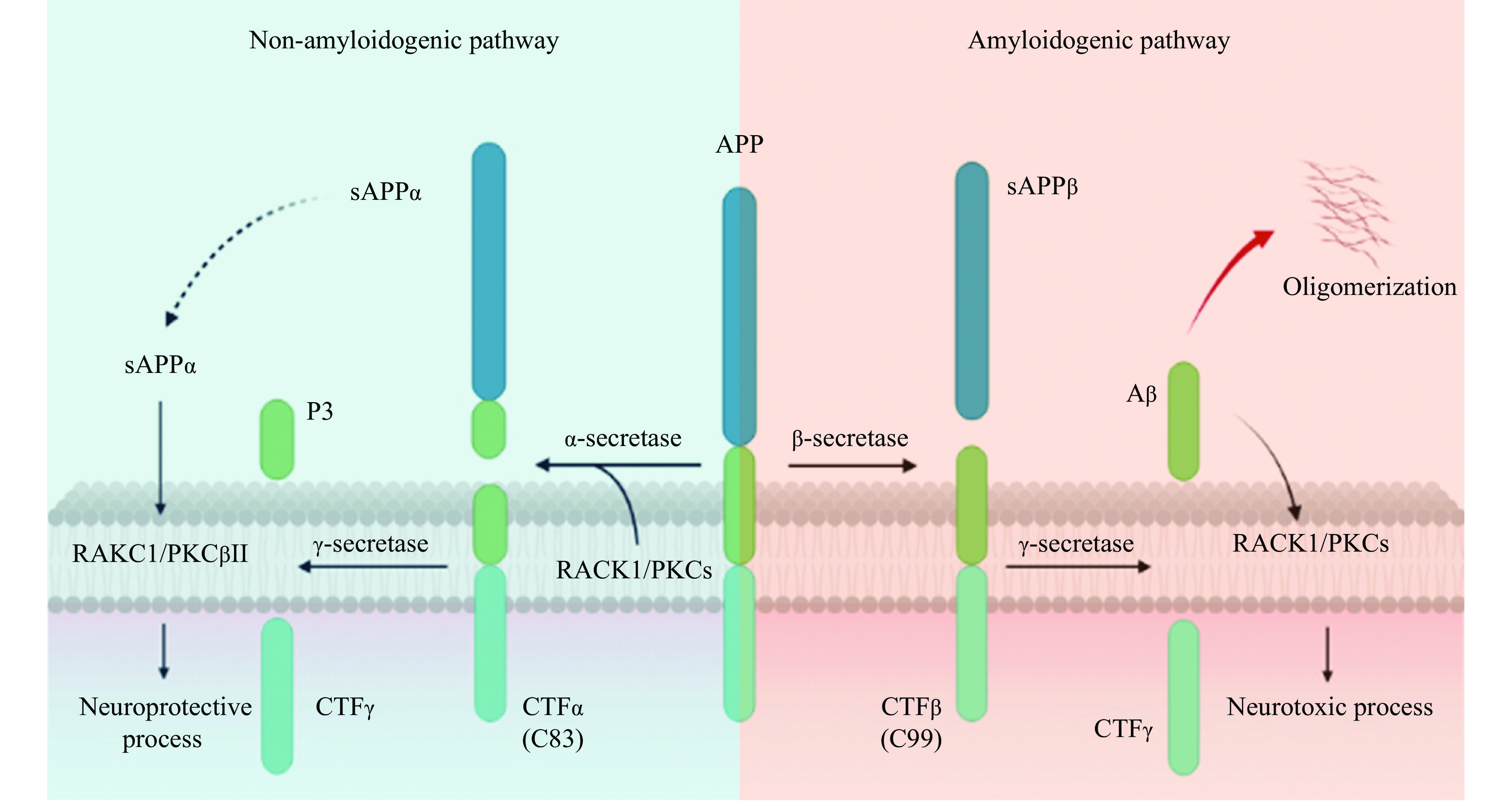
The receptor for activated C kinase 1 (RACK1) is a protein that plays a crucial role in various signaling pathways and is involved in the pathogenesis of Alzheimer's disease (AD), a prevalent neurodegenerative disease. RACK1 is highly expressed in neuronal cells of the central nervous system and regulates the pathogenesis of AD. Specifically, RACK1 is involved in regulation of the amyloid-β precursor protein processing through α- or β-secretase by binding to different protein kinase C isoforms. Additionally, RACK1 promotes synaptogenesis and synaptic plasticity by inhibiting N-methyl-D-aspartate receptors and activating gamma-aminobutyric acid A receptors, thereby preventing neuronal excitotoxicity. RACK1 also assembles inflammasomes that are involved in various neuroinflammatory pathways, such as nuclear factor-kappa B, tumor necrosis factor-alpha, and NOD-like receptor family pyrin domain-containing 3 pathways. The potential to design therapeutics that block amyloid-β accumulation and inflammation or precisely regulate synaptic plasticity represents an attractive therapeutic strategy, in which RACK1 is a potential target. In this review, we summarize the contribution of RACK1 to the pathogenesis of AD and its potential as a therapeutic target.
This work was supported by grants from the National Natural Science Foundation of China (Grant No. 82071395), the Natural Science Foundation of Chongqing (Grant Nos. cstc2021ycjh-bgzxm0186, cstc2020jcyj-zdxmX0004, and cstc2021jcyj-bsh0023) and the CQMU Program for Youth Innovation in Future Medicine (Grant No. W0044).
None.
CLC number: R749.16, Document code: A
The authors reported no conflict of interests.
| [1] |
Torrisi SA, Geraci F, Tropea MR, et al. Fluoxetine and vortioxetine reverse depressive-like phenotype and memory deficits induced by Aβ1–42 oligomers in mice: a key role of transforming growth factor-β1[J]. Front Pharmacol, 2019, 10: 693. doi: 10.3389/fphar.2019.00693
|
| [2] |
Scheltens P, De Strooper B, Kivipelto M, et al. Alzheimer's disease[J]. Lancet, 2021, 397(10284): 1577–1590. doi: 10.1016/S0140-6736(20)32205-4
|
| [3] |
Morgen K, Frölich L. The metabolism hypothesis of Alzheimer's disease: from the concept of central insulin resistance and associated consequences to insulin therapy[J]. J Neural Transm, 2015, 122(4): 499–504. doi: 10.1007/s00702-015-1377-5
|
| [4] |
Wang M, Miao D, Cao X, et al. Innate immune activation in Alzheimer's disease[J]. Ann Transl Med, 2018, 6(10): 177. doi: 10.21037/atm.2018.04.20
|
| [5] |
Sharma D, Kanneganti TD. The cell biology of inflammasomes: mechanisms of inflammasome activation and regulation[J]. J Cell Biol, 2016, 213(6): 617–629. doi: 10.1083/jcb.201602089
|
| [6] |
Battaini F, Pascale A, Paoletti R, et al. The role of anchoring protein RACK1 in PKC activation in the ageing rat brain[J]. Trends Neurosci, 1997, 20(9): 410–415. doi: 10.1016/S0166-2236(97)01084-9
|
| [7] |
Buoso E, Galasso M, Serafini MM, et al. Transcriptional regulation of RACK1 and modulation of its expression: role of steroid hormones and significance in health and aging[J]. Cell Signal, 2017, 35: 264–271. doi: 10.1016/j.cellsig.2017.02.010
|
| [8] |
Besson A, Wilson TL, Yong VW. The anchoring protein RACK1 links protein kinase Cε to integrin β chains. Requirement for adhesion and motility[J]. J Biol Chem, 2002, 277(24): 22073–22084. doi: 10.1074/jbc.M111644200
|
| [9] |
Slager RE, Devasure JM, Pavlik JA, et al. RACK1, a PKC targeting protein, is exclusively localized to basal airway epithelial cells[J]. J Histochem Cytochem, 2008, 56(1): 7–14. doi: 10.1369/jhc.7A7249.2007
|
| [10] |
Pass JM, Gao J, Jones WK, et al. Enhanced PKCβII translocation and PKCβII-RACK1 interactions in PKCε-induced heart failure: a role for RACK1[J]. Am J Physiol Heart Circ Physiol, 2001, 281(6): H2500–H2510. doi: 10.1152/ajpheart.2001.281.6.H2500
|
| [11] |
Grosso S, Volta V, Sala LA, et al. PKCβII modulates translation independently from mTOR and through RACK1[J]. Biochem J, 2008, 415(1): 77–85. doi: 10.1042/BJ20080463
|
| [12] |
Pass JM, Zheng Y, Wead WB, et al. PKCε activation induces dichotomous cardiac phenotypes and modulates PKCε-RACK interactions and RACK expression[J]. Am J Physiol Heart Circ Physiol, 2001, 280(3): H946–H955. doi: 10.1152/ajpheart.2001.280.3.H946
|
| [13] |
Schechtman D, Mochly-Rosen D. Adaptor proteins in protein kinase C-mediated signal transduction[J]. Oncogene, 2001, 20(44): 6339–6347. doi: 10.1038/sj.onc.1204778
|
| [14] |
Lei J, Li Q, Gao Y, et al. Increased PKCα activity by Rack1 overexpression is responsible for chemotherapy resistance in T-cell acute lymphoblastic leukemia-derived cell line[J]. Sci Rep, 2016, 6: 33717. doi: 10.1038/srep33717
|
| [15] |
Osmanagic-Myers S, Wiche G. Plectin-RACK1 (receptor for activated C kinase 1) scaffolding: a novel mechanism to regulate protein kinase C activity[J]. J Biol Chem, 2004, 279(18): 18701–18710. doi: 10.1074/jbc.M312382200
|
| [16] |
Chen YH, Wang HC, Lin CY, et al. Effects of prenyl pyrophosphates on the binding of PKCγ with RACK1[J]. J Exp Zool A Comp Exp Biol, 2003, 295A(1): 71–82. doi: 10.1002/jez.a.10213
|
| [17] |
Dell EJ, Connor J, Chen S, et al. The βγ subunit of heterotrimeric G proteins interacts with RACK1 and two other WD repeat proteins[J]. J Biol Chem, 2002, 277(51): 49888–49895. doi: 10.1074/jbc.M202755200
|
| [18] |
Patterson RL, van Rossum DB, Barrow RK, et al. RACK1 binds to inositol 1, 4, 5-trisphosphate receptors and mediates Ca2+ release[J]. Proc Natl Acad Sci U S A, 2004, 101(8): 2328–2332. doi: 10.1073/pnas.0308567100
|
| [19] |
Rodriguez MM, Ron D, Touhara K, et al. RACK1, a protein kinase C anchoring protein, coordinates the binding of activated protein kinase C and select pleckstrin homology domains in vitro[J]. Biochemistry, 1999, 38(42): 13787–13794. doi: 10.1021/bi991055k
|
| [20] |
Brandon NJ, Uren JM, Kittler JT, et al. Subunit-specific association of protein kinase C and the receptor for activated C kinase with GABA type A receptors[J]. J Neurosci, 1999, 19(21): 9228–9234. doi: 10.1523/JNEUROSCI.19-21-09228.1999
|
| [21] |
Yaka R, Thornton C, Vagts AJ, et al. NMDA receptor function is regulated by the inhibitory scaffolding protein, RACK1[J]. Proc Natl Acad Sci U S A, 2002, 99(8): 5710–5715. doi: 10.1073/pnas.062046299
|
| [22] |
Adams DR, Ron D, Kiely PA. RACK1, A multifaceted scaffolding protein: structure and function[J]. Cell Commun Signal, 2011, 9: 22. doi: 10.1186/1478-811X-9-22
|
| [23] |
Van der Zee EA, Palm IF, O'Connor M, et al. Aging-related alterations in the distribution of Ca2+-dependent PKC isoforms in rabbit hippocampus[J]. Hippocampus, 2004, 14(7): 849–860. doi: 10.1002/hipo.20000
|
| [24] |
Pascale A, Fortino I, Govoni S, et al. Functional impairment in protein kinase C by RACK1 (receptor for activated C kinase 1) deficiency in aged rat brain cortex[J]. J Neurochem, 1996, 67(6): 2471–2477. doi: 10.1046/j.1471-4159.1996.67062471.x
|
| [25] |
Peyrl A, Weitzdoerfer R, Gulesserian T, et al. Aberrant expression of signaling-related proteins 14-3-3 gamma and RACK1 in fetal down syndrome brain (trisomy 21)[J]. Electrophoresis, 2002, 23(1): 152–157. doi: 10.1002/1522-2683(200201)23:1<152::AID-ELPS152>3.0.CO;2-T
|
| [26] |
Alkon DL, Sun MK, Nelson TJ. PKC signaling deficits: a mechanistic hypothesis for the origins of Alzheimer's disease[J]. Trends Pharmacol Sci, 2007, 28(2): 51–60. doi: 10.1016/j.tips.2006.12.002
|
| [27] |
Ni H, Rui Q, Xu Y, et al. RACK1 upregulation induces neuroprotection by activating the IRE1-XBP1 signaling pathway following traumatic brain injury in rats[J]. Exp Neurol, 2018, 304: 102–113. doi: 10.1016/j.expneurol.2018.03.003
|
| [28] |
Tanzi RE, Bertram L. Twenty years of the Alzheimer's disease amyloid hypothesis: a genetic perspective[J]. Cell, 2005, 120(4): 545–555. doi: 10.1016/j.cell.2005.02.008
|
| [29] |
Zhu J, Chen X, Song Y, et al. Deficit of RACK1 contributes to the spatial memory impairment via upregulating BECLIN1 to induce autophagy[J]. Life Sci, 2016, 151: 115–121. doi: 10.1016/j.lfs.2016.02.014
|
| [30] |
Wang X, Zhou X, Li G, et al. Modifications and trafficking of APP in the pathogenesis of Alzheimer's disease[J]. Front Mol Neurosci, 2017, 10: 294. doi: 10.3389/fnmol.2017.00294
|
| [31] |
Kögel D, Deller T, Behl C. Roles of amyloid precursor protein family members in neuroprotection, stress signaling and aging[J]. Exp Brain Res, 2012, 217(3–4): 471–479. doi: 10.1007/s00221-011-2932-4
|
| [32] |
Turner PR, O'Connor K, Tate WP, et al. Roles of amyloid precursor protein and its fragments in regulating neural activity, plasticity and memory[J]. Prog Neurobiol, 2003, 70(1): 1–32. doi: 10.1016/S0301-0082(03)00089-3
|
| [33] |
Fol R, Braudeau J, Ludewig S, et al. Viral gene transfer of APPsα rescues synaptic failure in an Alzheimer's disease mouse model[J]. Acta Neuropathol, 2016, 131(2): 247–266. doi: 10.1007/s00401-015-1498-9
|
| [34] |
Hampel H, Hardy J, Blennow K, et al. The amyloid-β pathway in Alzheimer's disease[J]. Mol Psychiatry, 2021, 26(10): 5481–5503. doi: 10.1038/s41380-021-01249-0
|
| [35] |
Hardy J, Selkoe DJ. The amyloid hypothesis of Alzheimer's disease: progress and problems on the road to therapeutics[J]. Science, 2002, 297(5580): 353–356. doi: 10.1126/science.1072994
|
| [36] |
de Barry J, Liégeois CM, Janoshazi A. Protein kinase C as a peripheral biomarker for Alzheimer's disease[J]. Exp Gerontol, 2010, 45(1): 64–69. doi: 10.1016/j.exger.2009.10.015
|
| [37] |
Shimohama S, Kamiya S, Taniguchi T, et al. Intracellular receptors for activated C-kinase in the postmortem human brain: no alteration in Alzheimer disease[J]. Alzheimer Dis Assoc Disord, 1998, 12(4): 384–386. doi: 10.1097/00002093-199812000-00022
|
| [38] |
He W, Tu M, Du Y, et al. Nicotine promotes AβPP nonamyloidogenic processing via RACK1-dependent activation of PKC in SH-SY5Y-AβPP695 cells[J]. J Alzheimers Dis, 2020, 75(2): 451–460. doi: 10.3233/JAD-200003
|
| [39] |
Sato N, Shinohara M, Rakugi H, et al. Dual effects of statins on Aβ metabolism: upregulation of the degradation of APP-CTF and Aβ clearance[J]. Neurodegener Dis, 2012, 10(1-4): 305–308. doi: 10.1159/000334534
|
| [40] |
Buoso E, Biundo F, Lanni C, et al. Modulation of Rack-1/PKCβII signalling by soluble AβPPα in SH-SY5Y cells[J]. Curr Alzheimer Res, 2013, 10(7): 697–705. doi: 10.2174/15672050113109990145
|
| [41] |
Lee W, Boo JH, Jung MW, et al. Amyloid beta peptide directly inhibits PKC activation[J]. Mol Cell Neurosci, 2004, 26(2): 222–231. doi: 10.1016/j.mcn.2003.10.020
|
| [42] |
Cheng G, Yu Z, Zhou D, et al. Phosphatidylinositol-3-kinase-Akt kinase and p42/p44 mitogen-activated protein kinases mediate neurotrophic and excitoprotective actions of a secreted form of amyloid precursor protein[J]. Exp Neurol, 2002, 175(2): 407–414. doi: 10.1006/exnr.2002.7920
|
| [43] |
Mattson MP, Camandola S. NF-κB in neuronal plasticity and neurodegenerative disorders[J]. J Clin Invest, 2001, 107(3): 247–254. doi: 10.1172/JCI11916
|
| [44] |
Thal DR, Rüb U, Orantes M, et al. Phases of Aβ-deposition in the human brain and its relevance for the development of AD[J]. Neurology, 2002, 58(12): 1791–1800. doi: 10.1212/WNL.58.12.1791
|
| [45] |
Liu W, Dou F, Feng J, et al. RACK1 is involved in β-amyloid impairment of muscarinic regulation of GABAergic transmission[J]. Neurobiol Aging, 2011, 32(10): 1818–1826. doi: 10.1016/j.neurobiolaging.2009.10.017
|
| [46] |
Saito M, Iwawaki T, Taya C, et al. Diphtheria toxin receptor-mediated conditional and targeted cell ablation in transgenic mice[J]. Nat Biotechnol, 2001, 19(8): 746–750. doi: 10.1038/90795
|
| [47] |
Favit A, Grimaldi M, Nelson TJ, et al. Alzheimer's-specific effects of soluble β-amyloid on protein kinase C-α and –γ degradation in human fibroblasts[J]. Proc Nat Acad Sci U S A, 1998, 95(10): 5562–5567. doi: 10.1073/pnas.95.10.5562
|
| [48] |
Choi DS, Wang D, Yu G, et al. PKCε increases endothelin converting enzyme activity and reduces amyloid plaque pathology in transgenic mice[J]. Proc Natl Acad Sci U S A, 2006, 103(21): 8215–8220. doi: 10.1073/pnas.0509725103
|
| [49] |
Zhu G, Wang D, Lin YH, et al. Protein kinase Cϵ suppresses Aβ production and promotes activation of α-secretase[J]. Biochem Biophys Res Commun, 2001, 285(4): 997–1006. doi: 10.1006/bbrc.2001.5273
|
| [50] |
Du Y, Zhao Y, Li C, et al. Inhibition of PKCδ reduces amyloid-β levels and reverses Alzheimer disease phenotypes[J]. J Exp Med, 2018, 215(6): 1665–1677. doi: 10.1084/jem.20171193
|
| [51] |
Sajan MP, Hansen BC, Higgs MG, et al. Atypical PKC, PKCλ/ι, activates β-secretase and increases Aβ1–40/42 and phospho-tau in mouse brain and isolated neuronal cells, and may link hyperinsulinemia and other aPKC activators to development of pathological and memory abnormalities in Alzheimer's disease[J]. Neurobiol Aging, 2018, 61: 225–237. doi: 10.1016/j.neurobiolaging.2017.09.001
|
| [52] |
Mazanetz MP, Fischer PM. Untangling tau hyperphosphorylation in drug design for neurodegenerative diseases[J]. Nat Rev Drug Discov, 2007, 6(6): 464–479. doi: 10.1038/nrd2111
|
| [53] |
Takashima A. GSK-3 is essential in the pathogenesis of Alzheimer's disease[J]. J Alzheimers Dis, 2006, 9(S3): 309–317. doi: 10.3233/JAD-2006-9S335
|
| [54] |
Hanseeuw BJ, Betensky RA, Jacobs HIL, et al. Association of amyloid and tau with cognition in preclinical alzheimer disease: a longitudinal study[J]. JAMA Neurol, 2019, 76(8): 915–924. doi: 10.1001/jamaneurol.2019.1424
|
| [55] |
Isagawa T, Mukai H, Oishi K, et al. Dual effects of PKNα and protein kinase C on phosphorylation of tau protein by glycogen synthase kinase-3β[J]. Biochem Biophys Res Commun, 2000, 273(1): 209–212. doi: 10.1006/bbrc.2000.2926
|
| [56] |
Zhang D, Wang Q, Zhu T, et al. RACK1 promotes the proliferation of THP1 acute myeloid leukemia cells[J]. Mol Cell Biochem, 2013, 384(1–2): 197–202. doi: 10.1007/s11010-013-1798-0
|
| [57] |
Collingridge GL, Isaac JTR, Wang Y. Receptor trafficking and synaptic plasticity[J]. Nat Rev Neurosci, 2004, 5(12): 952–962. doi: 10.1038/nrn1556
|
| [58] |
Lau CG, Takeuchi K, Rodenas-Ruano A, et al. Regulation of NMDA receptor Ca2+ signalling and synaptic plasticity[J]. Biochem Soc Trans, 2009, 37(Pt 6): 1369–1374.
|
| [59] |
Hansen KB, Yi F, Perszyk RE, et al. Structure, function, and allosteric modulation of NMDA receptors[J]. J Gen Physiol, 2018, 150(8): 1081–1105. doi: 10.1085/jgp.201812032
|
| [60] |
Petit-Pedrol M, Groc L. Regulation of membrane NMDA receptors by dynamics and protein interactions[J]. J Cell Biol, 2021, 220(1): e202006101. doi: 10.1083/jcb.202006101
|
| [61] |
Kojima N, Wang J, Mansuy IM, et al. Rescuing impairment of long-term potentiation in fyn-deficient mice by introducing Fyn transgene[J]. Proc Natl Acad Sci U S A, 1997, 94(9): 4761–4765. doi: 10.1073/pnas.94.9.4761
|
| [62] |
Thornton C, Tang KC, Phamluong K, et al. Spatial and temporal regulation of RACK1 function and N-methyl-D-aspartate receptor activity through WD40 motif-mediated dimerization[J]. J Biol Chem, 2004, 279(30): 31357–31364. doi: 10.1074/jbc.M402316200
|
| [63] |
Yaka R, Phamluong K, Ron D. Scaffolding of Fyn kinase to the NMDA receptor determines brain region sensitivity to ethanol[J]. J Neurosci, 2003, 23(9): 3623–3632. doi: 10.1523/JNEUROSCI.23-09-03623.2003
|
| [64] |
Wang J, Carnicella S, Phamluong K, et al. Ethanol induces long-term facilitation of NR2B-NMDA receptor activity in the dorsal striatum: implications for alcohol drinking behavior[J]. J Neurosci, 2007, 27(13): 3593–3602. doi: 10.1523/JNEUROSCI.4749-06.2007
|
| [65] |
Phamluong K, Darcq E, Wu S, et al. Fyn signaling is compartmentalized to dopamine D1 receptor expressing neurons in the dorsal medial striatum[J]. Front Mol Neurosci, 2017, 10: 273. doi: 10.3389/fnmol.2017.00273
|
| [66] |
Raymond CR, Redman SJ. Different calcium sources are narrowly tuned to the induction of different forms of LTP[J]. J Neurophysiol, 2002, 88(1): 249–255. doi: 10.1152/jn.2002.88.1.249
|
| [67] |
Ambrad Giovannetti E, Fuhrmann M. Unsupervised excitation: GABAergic dysfunctions in Alzheimer's disease[J]. Brain Res, 2019, 1707: 216–226. doi: 10.1016/j.brainres.2018.11.042
|
| [68] |
Palop JJ, Mucke L. Network abnormalities and interneuron dysfunction in Alzheimer disease[J]. Nat Rev Neurosci, 2016, 17(12): 777–792. doi: 10.1038/nrn.2016.141
|
| [69] |
Samakashvili S, Ibáñez C, Simó C, et al. Analysis of chiral amino acids in cerebrospinal fluid samples linked to different stages of Alzheimer disease[J]. Electrophoresis, 2011, 32(19): 2757–2764. doi: 10.1002/elps.201100139
|
| [70] |
Yoshiike Y, Kimura T, Yamashita S, et al. GABAA receptor-mediated acceleration of aging-associated memory decline in APP/PS1 mice and its pharmacological treatment by picrotoxin[J]. PLoS One, 2008, 3(8): e3029. doi: 10.1371/journal.pone.0003029
|
| [71] |
Brandon NJ, Jovanovic JN, Smart TG, et al. Receptor for activated C kinase-1 facilitates protein kinase C-dependent phosphorylation and functional modulation of GABAA receptors with the activation of G-protein-coupled receptors[J]. J Neurosci, 2002, 22(15): 6353–6361. doi: 10.1523/JNEUROSCI.22-15-06353.2002
|
| [72] |
Meier J, Akyeli J, Kirischuk S, et al. GABAA receptor activity and PKC control inhibitory synaptogenesis in CNS tissue slices[J]. Mol Cell Neurosci, 2003, 23(4): 600–613. doi: 10.1016/S1044-7431(03)00079-4
|
| [73] |
Feng J, Cai X, Zhao J, et al. Serotonin receptors modulate GABAA receptor channels through activation of anchored protein kinase C in prefrontal cortical neurons[J]. J Neurosci, 2001, 21(17): 6502–6511. doi: 10.1523/JNEUROSCI.21-17-06502.2001
|
| [74] |
Littlejohn EL, Boychuk CR. Protein kinase C-dependent effects of neurosteroids on synaptic GABAA receptor inhibition require the δ-subunit[J]. Front Physiol, 2021, 12: 742838. doi: 10.3389/fphys.2021.742838
|
| [75] |
Chen J, He Y, Wu Y, et al. Single ethanol withdrawal regulates extrasynaptic δ-GABAA receptors via PKCδ activation[J]. Front Mol Neurosci, 2018, 11: 141. doi: 10.3389/fnmol.2018.00141
|
| [76] |
Li Q, Li Q, Jia J, et al. Sodium valproate ameliorates neuronal apoptosis in a kainic acid model of epilepsy via enhancing PKC-dependent GABAAR γ2 serine 327 phosphorylation[J]. Neurochem Res, 2018, 43(12): 2343–2352. doi: 10.1007/s11064-018-2659-8
|
| [77] |
Weerasinghe-Mudiyanselage PDE, Ang MJ, Kang S, et al. Structural plasticity of the hippocampus in neurodegenerative diseases[J]. Int J Mol Sci, 2022, 23(6): 3349. doi: 10.3390/ijms23063349
|
| [78] |
Mufson EJ, Mahady L, Waters D, et al. Hippocampal plasticity during the progression of Alzheimer's disease[J]. Neuroscience, 2015, 309: 51–67. doi: 10.1016/j.neuroscience.2015.03.006
|
| [79] |
Kershner L, Welshhans K. RACK1 is necessary for the formation of point contacts and regulates axon growth[J]. Dev Neurobiol, 2017, 77(9): 1038–1056. doi: 10.1002/dneu.22491
|
| [80] |
Kershner L, Welshhans K. RACK1 regulates neural development[J]. Neural Regen Res, 2017, 12(7): 1036–1039. doi: 10.4103/1673-5374.211175
|
| [81] |
Sklan EH, Podoly E, Soreq H. RACK1 has the nerve to act: structure meets function in the nervous system[J]. Prog Neurobiol, 2006, 78(2): 117–134. doi: 10.1016/j.pneurobio.2005.12.002
|
| [82] |
Yang H, Yang C, Zhu Q, et al. Rack1 controls parallel fiber–purkinje cell synaptogenesis and synaptic transmission[J]. Front Cell Neurosci, 2019, 13: 539. doi: 10.3389/fncel.2019.00539
|
| [83] |
Li H, Shang J, Zhang C, et al. Repetitive transcranial magnetic stimulation alleviates neurological deficits after cerebral ischemia through interaction between RACK1 and BDNF exon IV by the phosphorylation-dependent factor MeCP2[J]. Neurotherapeutics, 2020, 17(2): 651–663. doi: 10.1007/s13311-019-00771-y
|
| [84] |
Ceci M, Welshhans K, Ciotti MT, et al. RACK1 is a ribosome scaffold protein for β-actin mRNA/ZBP1 complex[J]. PLoS One, 2012, 7(4): e35034. doi: 10.1371/journal.pone.0035034
|
| [85] |
Dwane S, Durack E, O'Connor R, et al. RACK1 promotes neurite outgrowth by scaffolding AGAP2 to FAK[J]. Cell Signal, 2014, 26(1): 9–18. doi: 10.1016/j.cellsig.2013.08.036
|
| [86] |
Ju Hwang C, Choi DY, Park MH, et al. NF-κB as a key mediator of brain inflammation in Alzheimer's disease[J]. CNS Neurol Disord Drug Targets, 2019, 18(1): 3–10. doi: 10.2174/1871527316666170807130011
|
| [87] |
Heppner FL, Ransohoff RM, Becher B. Immune attack: the role of inflammation in Alzheimer disease[J]. Nat Rev Neurosci, 2015, 16(6): 358–372. doi: 10.1038/nrn3880
|
| [88] |
Morgan MJ, Liu Z. Crosstalk of reactive oxygen species and NF-κB signaling[J]. Cell Res, 2011, 21(1): 103–115. doi: 10.1038/cr.2010.178
|
| [89] |
Chami L, Buggia-Prévot V, Duplan E, et al. Nuclear factor-κB regulates βAPP and β- and γ-secretases differently at physiological and supraphysiological Aβ concentrations[J]. J Biol Chem, 2012, 287(29): 24573–24584. doi: 10.1074/jbc.M111.333054
|
| [90] |
Waiskopf N, Ofek K, Gilboa-Geffen A, et al. AChE and RACK1 promote the anti-inflammatory properties of fluoxetine[J]. J Mol Neurosci, 2014, 53(3): 306–315. doi: 10.1007/s12031-013-0174-6
|
| [91] |
Birikh KR, Sklan EH, Shoham S, et al. Interaction of "readthrough" acetylcholinesterase with RACK1 and PKCβⅡ correlates with intensified fear-induced conflict behavior[J]. Proc Natl Acad Sci U S A, 2003, 100(1): 283–288. doi: 10.1073/pnas.0135647100
|
| [92] |
Nijholt I, Farchi N, Kye M, et al. Stress-induced alternative splicing of acetylcholinesterase results in enhanced fear memory and long-term potentiation[J]. Mol Psychiatry, 2004, 9(2): 174–183. doi: 10.1038/sj.mp.4001446
|
| [93] |
Yin H, Song S, Pan X. Knockdown of miR-155 protects microglia against LPS-induced inflammatory injury via targeting RACK1: a novel research for intracranial infection[J]. J Inflamm, 2017, 14: 17. doi: 10.1186/s12950-017-0162-7
|
| [94] |
Teng Y, Zhang M, Wang W, et al. Compound danshen tablet ameliorated aβ25–35-induced spatial memory impairment in mice via rescuing imbalance between cytokines and neurotrophins[J]. BMC Complement Altern Med, 2014, 14: 23. doi: 10.1186/1472-6882-14-23
|
| [95] |
Chou W, Guo Z, Guo H, et al. AIM2 in regulatory T cells restrains autoimmune diseases[J]. Nature, 2021, 591(7849): 300–305. doi: 10.1038/s41586-021-03231-w
|
| [96] |
Wang Q, Zhou S, Wang J, et al. RACK1 antagonizes TNF-α-induced cell death by promoting p38 activation[J]. Sci Rep, 2015, 5: 14298. doi: 10.1038/srep14298
|
| [97] |
Duan Y, Zhang L, Angosto-Bazarra D, et al. RACK1 mediates NLRP3 inflammasome activation by promoting NLRP3 active conformation and inflammasome assembly[J]. Cell Rep, 2020, 33(7): 108405. doi: 10.1016/j.celrep.2020.108405
|
| [98] |
Buoso E, Masi M, Galbiati V, et al. Effect of estrogen-active compounds on the expression of RACK1 and immunological implications[J]. Arch Toxicol, 2020, 94(6): 2081–2095. doi: 10.1007/s00204-020-02756-9
|
| [99] |
Khan TK, Alkon DL. An internally controlled peripheral biomarker for Alzheimer's disease: Erk1 and Erk2 responses to the inflammatory signal bradykinin[J]. Proc Natl Acad Sci U S A, 2006, 103(35): 13203–13207. doi: 10.1073/pnas.0605411103
|
| [100] |
Heneka MT, Kummer MP, Stutz A, et al. NLRP3 is activated in Alzheimer's disease and contributes to pathology in APP/PS1 mice[J]. Nature, 2013, 493(7434): 674–678. doi: 10.1038/nature11729
|
| [101] |
Qu Z, Zhou J, Zhou Y, et al. Mycobacterial EST12 activates a RACK1-NLRP3-gasdermin D pyroptosis-IL-1β immune pathway[J]. Sci Adv, 2020, 6(43): eaba4733. doi: 10.1126/sciadv.aba4733
|
| [102] |
Nayak D, Roth TL, McGavern DB. Microglia development and function[J]. Annu Rev Immunol, 2014, 32: 367–402. doi: 10.1146/annurev-immunol-032713-120240
|
| [103] |
Wang Y, Ulland TK, Ulrich JD, et al. TREM2-mediated early microglial response limits diffusion and toxicity of amyloid plaques[J]. J Exp Med, 2016, 213(5): 667–675. doi: 10.1084/jem.20151948
|
| [104] |
Yin H, Song S, Pan X. Correction to: knockdown of miR-155 protects microglia against LPS-induced inflammatory injury via targeting RACK1: a novel research for intracranial infection[J]. J Inflamm, 2018, 15: 6. doi: 10.1186/s12950-018-0183-x
|
| [105] |
Dan H, Liu S, Liu J, et al. RACK1 promotes cancer progression by increasing the M2/M1 macrophage ratio via the NF-κB pathway in oral squamous cell carcinoma[J]. Mol Oncol, 2020, 14(4): 795–807. doi: 10.1002/1878-0261.12644
|
| [106] |
Sun Z, Tang X, Lin F, et al. The WD40 repeat protein WDR26 binds Gβγ and promotes Gβγ-dependent signal transduction and leukocyte migration[J]. J Biol Chem, 2011, 286(51): 43902–43912. doi: 10.1074/jbc.M111.301382
|
| [107] |
Rigas AC, Ozanne DM, Neal DE, et al. The scaffolding protein RACK1 interacts with androgen receptor and promotes cross-talk through a protein kinase C signaling pathway[J]. J Biol Chem, 2003, 278(46): 46087–46093. doi: 10.1074/jbc.M306219200
|
| [108] |
Corsini E, Galbiati V, Papale A, et al. Role of androgens in dhea-induced rack1 expression and cytokine modulation in monocytes[J]. Immun Ageing, 2016, 13: 20. doi: 10.1186/s12979-016-0075-y
|
| [109] |
Corsini E, Lucchi L, Meroni M, et al. In vivo dehydroepiandrosterone restores age-associated defects in the protein kinase C signal transduction pathway and related functional responses[J]. J Immunol, 2002, 168(4): 1753–1758. doi: 10.4049/jimmunol.168.4.1753
|
| [110] |
Arnold JT. DHEA metabolism in prostate: for better or worse?[J]. Mol Cell Endocrinol, 2009, 301(1–2): 83–88. doi: 10.1016/j.mce.2008.10.019
|
| [111] |
Pinto A, Malacrida B, Oieni J, et al. DHEA modulates the effect of cortisol on RACK1 expression via interference with the splicing of the glucocorticoid receptor[J]. Br J Pharmacol, 2015, 172(11): 2918–2927. doi: 10.1111/bph.13097
|
| [112] |
Li X, Li J, Qian J, et al. Loss of ribosomal RACK1 (receptor for activated protein kinase C 1) induced by phosphorylation at T50 alleviates cerebral ischemia-reperfusion injury in rats[J]. Stroke, 2019, 50(1): 162–171. doi: 10.1161/STROKEAHA.118.022404
|
| [113] |
Zhao Y, Wang Q, Qiu G, et al. RACK1 promotes autophagy by enhancing the atg14L-beclin 1-Vps34-Vps15 complex formation upon phosphorylation by AMPK[J]. Cell Rep, 2015, 13(7): 1407–1417. doi: 10.1016/j.celrep.2015.10.011
|
| [114] |
Cao J, Zhao M, Liu J, et al. RACK1 promotes self-renewal and chemoresistance of cancer stem cells in human hepatocellular carcinoma through stabilizing nanog[J]. Theranostics, 2019, 9(3): 811–828. doi: 10.7150/thno.29271
|
| [115] |
Ashique AM, Kharazia V, Yaka R, et al. Localization of the scaffolding protein RACK1 in the developing and adult mouse brain[J]. Brain Res, 2006, 1069(1): 31–38. doi: 10.1016/j.brainres.2005.11.018
|
| [116] |
Bolger GB, Smoot LHM, van Groen T. Dominant-negative attenuation of cAMP-selective phosphodiesterase PDE4D action affects learning and behavior[J]. Int J Mol Sci, 2020, 21(16): 5704. doi: 10.3390/ijms21165704
|
| [117] |
Yarwood SJ, Parnell E, Bird RJ. The cyclic AMP phosphodiesterase 4D5 (PDE4D5)/receptor for activated C-kinase 1 (RACK1) signalling complex as a sensor of the extracellular nano-environment[J]. Cell Signall, 2017, 35: 282–289. doi: 10.1016/j.cellsig.2017.01.013
|
| [1] | Anastasia V. Poznyak, Alexey Aleksandrovich Yakovlev, Mikhail А. Popov, Alexander D. Zhuravlev, Vasily N. Sukhorukov, Alexander N. Orekhov. Coronary atherosclerotic plaque regression strategies[J]. The Journal of Biomedical Research. DOI: 10.7555/JBR.37.20230223 |
| [2] | Yao Daokuo, Gao Xiangyu, Zhao Huiqiang, Chen Hui, Wang Lexin. Multivessel coronary artery ectasia and severe calcification in a patient with pheochromocytoma: a case report[J]. The Journal of Biomedical Research, 2019, 33(1): 69-72. DOI: 10.7555/JBR.32.20170047 |
| [3] | Ling Yan, Shuang Ding, Bing Gu, Ping Ma. Clinical application of simultaneous detection of cystatin C, cathepsin S, and IL-1 in classification of coronary artery disease[J]. The Journal of Biomedical Research, 2017, 31(4): 315-320. DOI: 10.7555/JBR.31.20150152 |
| [4] | Jiawei Liao, Wei Huang, George Liu. Animal models of coronary heart disease[J]. The Journal of Biomedical Research, 2017, 31(1): 3-10. DOI: 10.7555/JBR.30.20150051 |
| [5] | Bernardo L Trigatti, Mark Fuller. HDL signaling and protection against coronary artery atherosclerosis in mice[J]. The Journal of Biomedical Research, 2016, 30(2): 94-100. DOI: 10.7555/JBR.30.20150079 |
| [6] | Lei Jiang, Yingfeng Tu, Hongcheng Shi, Zhen Cheng. PET probes beyond 18F-FDG[J]. The Journal of Biomedical Research, 2014, 28(6): 435-446. DOI: 10.7555/JBR.28.20130196 |
| [7] | Rajiv Shrestha, Jing Xu, Dujiang Xie, Zhizhong Liu, Tian Xu, Fei Ye, Shiqing Din, Xuesong Qian, Song Yang, Yueqiang Liu, Feng Li, Aiping Zhang, Shaoliang Chen. Comparison of clinical outcomes of Chinese men and women after coronary stenting for coronary artery disease: a multi-center retrospective analysis of 4,334 patients[J]. The Journal of Biomedical Research, 2014, 28(5): 368-375. DOI: 10.7555/JBR.28.20120127 |
| [8] | Min Zhang, Yan Zhang, Shuaishuai Zhu, Xiaoyu Li, Qing Yang, Hui Bai, Qi Chen. Genetic variants of the class A scavenger receptor gene are associated with coronary artery disease in Chinese[J]. The Journal of Biomedical Research, 2012, 26(6): 418-424. DOI: 10.7555/JBR.26.20110116 |
| [9] | Chunjian Li, Zhijian Yang, Kejiang Cao. Retrieval of dislodged coronary stent from left renal artery by gooseneck snare[J]. The Journal of Biomedical Research, 2010, 24(6): 479-482. DOI: 10.1016/S1674-8301(10)60064-4 |
| [10] | Yangyang Zhang, Yanhu Wu, Biao Yuan, Xiang Liu, Sheng Zhao, Zhi Li, Yu Xia. Coronary artery bypass grafting with concomitant resection for carcinoma of lung[J]. The Journal of Biomedical Research, 2010, 24(1): 77-80. |
Figures(2)
Supported by: Beijing Renhe Information Technology Co., Ltd. E-mail:
info@rhhz.net 
 Authors and Reviewers

 DownLoad: