Figures of the Article
-
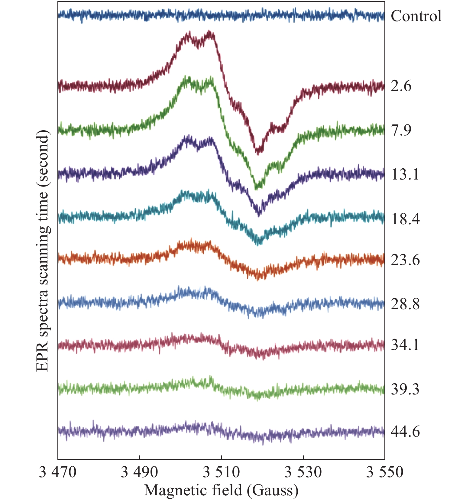
![]() Consecutively measured spectra of 340 μmol/L metHb (by haem) reacting with 1360 mmol/L H2O2 (a 4-fold excess of peroxide over haem).
Consecutively measured spectra of 340 μmol/L metHb (by haem) reacting with 1360 mmol/L H2O2 (a 4-fold excess of peroxide over haem).
-
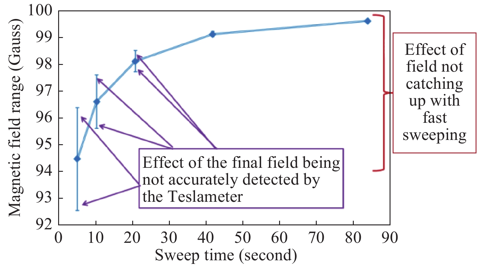
![]() Magnetic field range of a spectrum as reported by the Teslameter at different spectra scan rates.
Magnetic field range of a spectrum as reported by the Teslameter at different spectra scan rates.
-
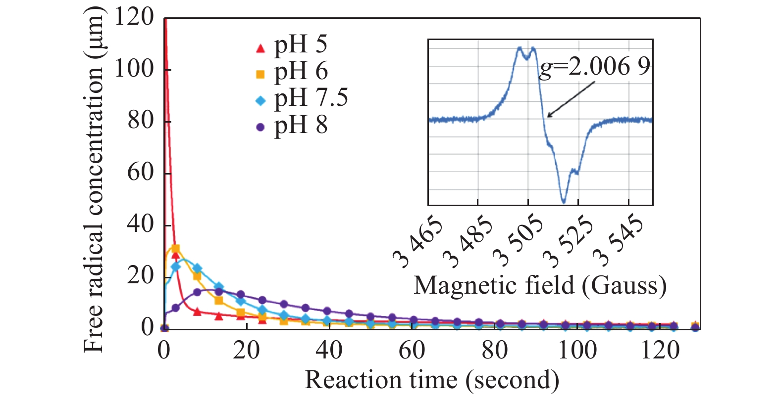
![]() Kinetic dependences of the tyrosyl free radical in the mixtures of 340 μmol/L human metHb and 1.36 mmol/L H2O2 (both concentrations are final), at different pH values.
Kinetic dependences of the tyrosyl free radical in the mixtures of 340 μmol/L human metHb and 1.36 mmol/L H2O2 (both concentrations are final), at different pH values.
-
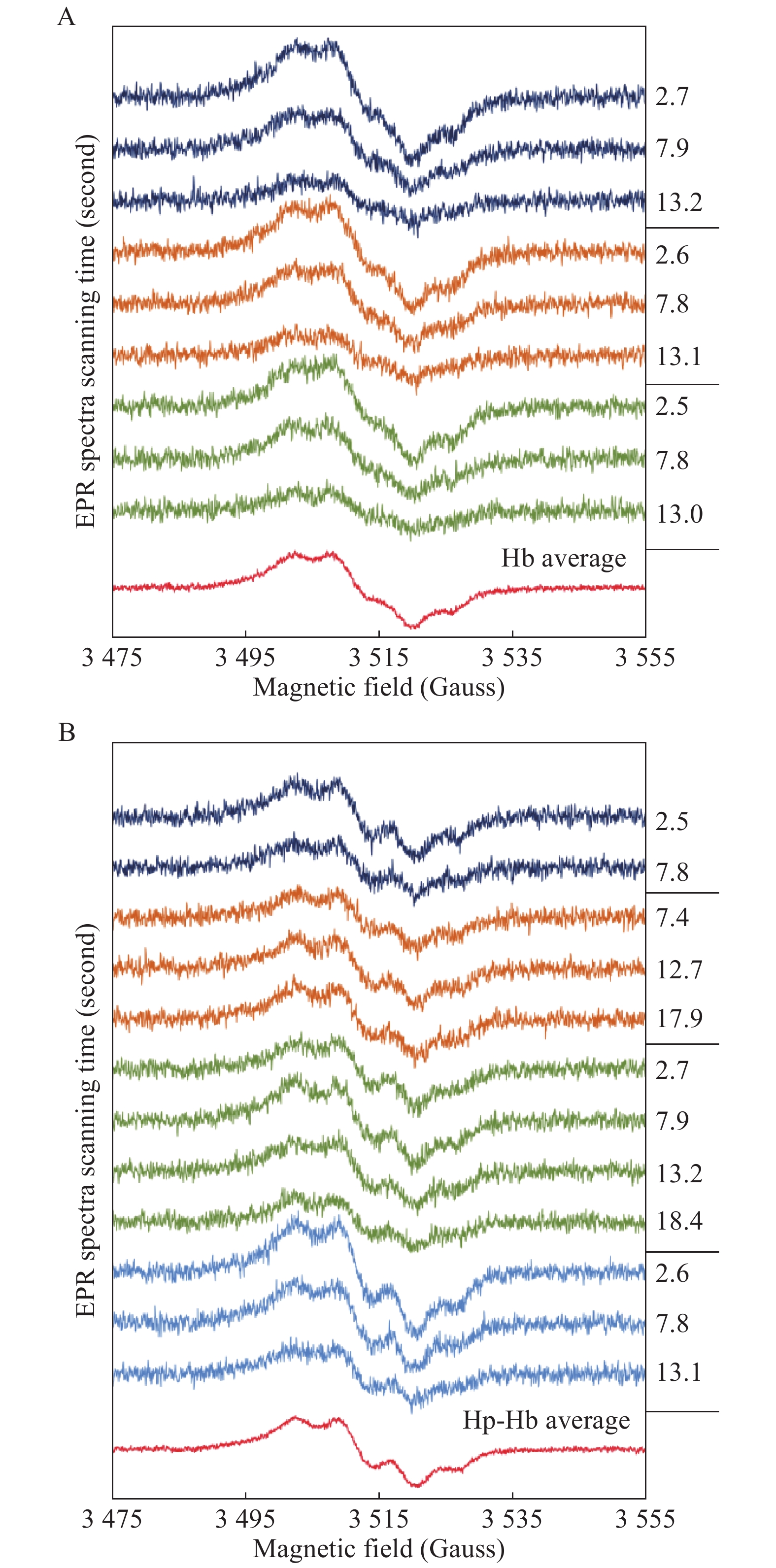
![]() Liquid phase EPR spectra of Tyr radical in Hb and Hp-Hb.
Liquid phase EPR spectra of Tyr radical in Hb and Hp-Hb.
-
![]() Kinetic dependences of the tyrosyl radical in the Hb+H2O2 and Hp-Hb+H2O2 systems.
Kinetic dependences of the tyrosyl radical in the Hb+H2O2 and Hp-Hb+H2O2 systems.
-
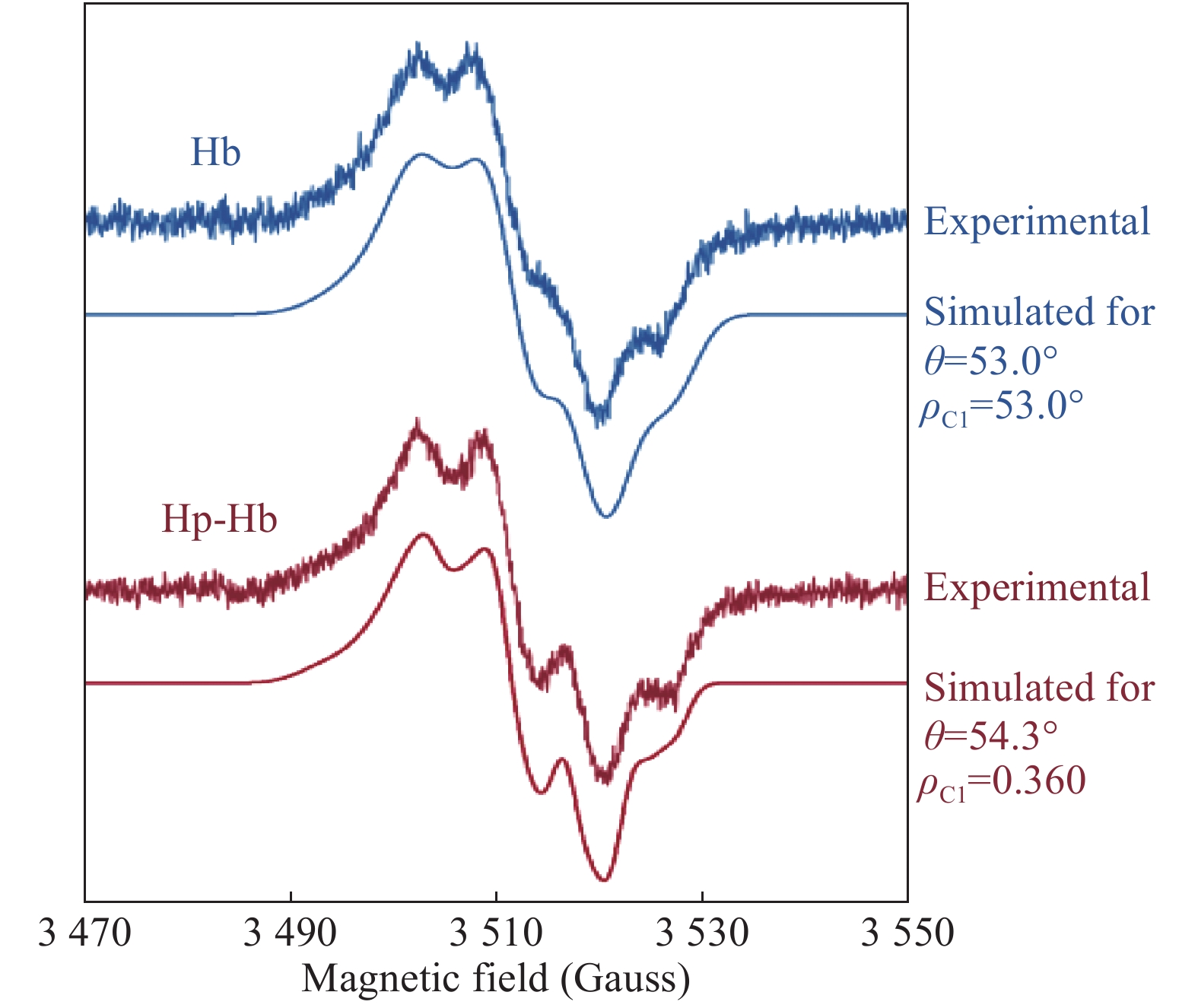
![]() The averaged EPR spectra of the free radicals formed under H2O2 addition to Hb and to the Hp-Hb complex (the same as shown in Fig. 4
) and their simulations as tyrosyl radical EPR signals.
The averaged EPR spectra of the free radicals formed under H2O2 addition to Hb and to the Hp-Hb complex (the same as shown in Fig. 4
) and their simulations as tyrosyl radical EPR signals.
-
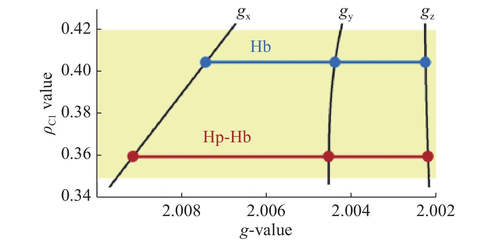
![]() A ρ-g chart[18,33] showing the principal g-factor sets for the tyrosyl radicals in Hb and Hp-Hb.
A ρ-g chart[18,33] showing the principal g-factor sets for the tyrosyl radicals in Hb and Hp-Hb.
-
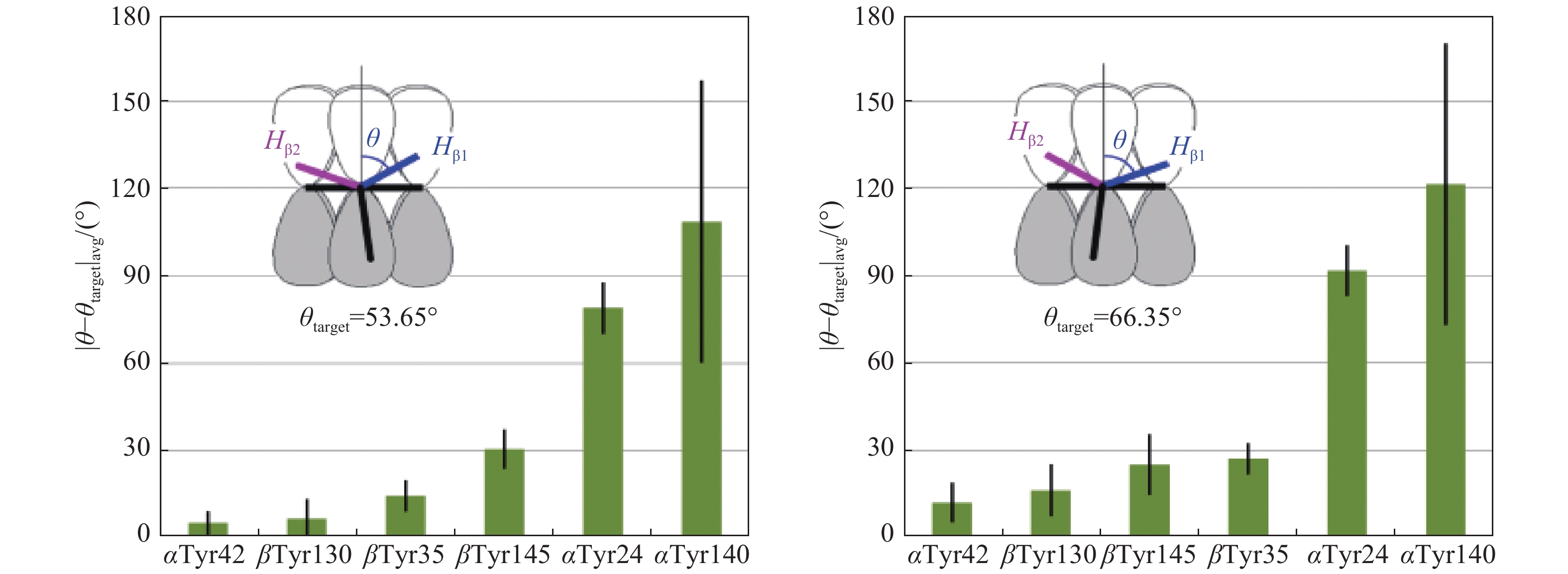
![]() The closeness of the phenol ring rotation angle θ in the six tyrosines in Hb to the target value of either 53.65° (average of the 53° and 54.3° angles reported in Fig. 6
) or its complementary angle of 66.35° (see Materials and methods).
The closeness of the phenol ring rotation angle θ in the six tyrosines in Hb to the target value of either 53.65° (average of the 53° and 54.3° angles reported in Fig. 6
) or its complementary angle of 66.35° (see Materials and methods).



 Authors and Reviewers
